Do you remember the moment where the sign across the street appeared blurry to you? Did you force yourself to squint so hard just so you can read what the words were saying? If so, you should probably make an appointment for an eye exam ASAP!
So, what can you expect in an eye exam?
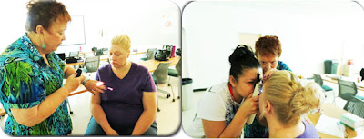
Students from the Ophthalmic Assistant Program at the Maricopa Skill Center demonstrate the process of a preliminary eye exam that is completed before meeting with an ophthalmologist.
 |
| Kerotometer in the Ophthalmic classroom at the Maricopa Skill Center. |
Medical History
The patient is asked if there are any known medical histories that could include complaints, allergic reactions, medications, diabetes, injuries, and surgeries.
Visual Acuity Test
This test is done to measure the sharpness of the patient's vision. He/she holds the occluder to cover one eye at a time while reading a Snellen chart or the near vision card.
 |
| MSC students assist one another through a preliminary eye exam. |
The patient is tested to see if he/she is able to see if they have peripheral vision around their visual field. This tests for any early signs of glaucoma, which is an eye disease where the nerve in the back of the eye is damaged.
Extraocular Muscles
This test examines the patient's eye muscles, which is identified as extraocular muscles. This is done using two fingers to make an "H" pattern to check that all muscles are intact.
Extraocular Muscles
This test examines the patient's eye muscles, which is identified as extraocular muscles. This is done using two fingers to make an "H" pattern to check that all muscles are intact.
Pupil Constriction
A pen light is directed towards the patient's eyes to check if it shrinks (constriction). The light is pointed directly at one eye at a time to see if the other eye reacts equally. When light is flashed at one eye, both pupils should constrict.
Other Eye Tests
A pen light is directed towards the patient's eyes to check if it shrinks (constriction). The light is pointed directly at one eye at a time to see if the other eye reacts equally. When light is flashed at one eye, both pupils should constrict.
 |
| Instructor Pat Lamb (left) shows how a direct ophthalmoscope is used. |
Other Eye Tests
There are other tests that are done while using different instruments to examine the inner eye. Instruments include a tonometer, a direct ophthalmoscope, or a slit lamp that's mounted on an exam chair.
Tonometer
This is used to measure inocular pressure, which is the inside pressure of the eyes. If there is pressure it may indicate the beginning causes of glaucoma.
Direct Ophthalmoscope
This is a magnified tool that is used to examine the retina, which is a thin tissue layer of the back of the eye. It has a mirror where a blue light reflects into the eye.
Hand-held Slit Lamp
This lamp is also known as a biomicroscope. Before this test is done, orange drops with fluorescein are put into the white area of the eye to indicate any scratches in the surface of the eye. This examines the cornea, iris, pupil, lens, lashes, and lids.
As you can see, getting an eye exam simply doesn't mean you're going in to get prescription eyeglasses or contact lens. Your eyes are being checked for eye disease and overall health!
Stay tuned for next week's program spotlight: Meat Cutting!


No comments:
Post a Comment